Foot digitization consists of capturing the imprint of the feet using various devices to establish an accurate diagnosis. This technique is particularly useful when it comes to designing custom-made plantar orthotics.
2D impressions are taken using a podograph. A kind of pressure-sensitive cushion, a scan or a mirror, the podograph detects and shows a perfect image of the foot’s plantar surface.
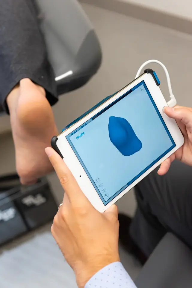
On the other hand, the 3D scanning method requires the contribution of a three-dimensional sensor. It can accurately reproduce the entire morphology of the foot.
These scanning methods make it possible to conceptualize the foot and determine the source of discomfort extremely rapidly.
Why take a foot imprint?
For the podiatrist, taking an imprint is both a diagnostic measure and a solution for creating highly sophisticated foot orthotics.
In addition to creating high-precision orthotics, foot scanning plays a key role in understanding postural imbalances.
The 2D technology acts as a sensor and provides a detailed mapping of the pressure points under the foot.
As a general rule, a two-dimensional scan highlights the following plantar characteristics:
- The size of the foot
- The type of arch of the plantar arch (flat foot, hollow foot or medium foot)
- The position of the foot when it is flat
- Weight distribution on the underside of the foot
Although faster than the traditional plaster casting method, digitizing the sole of the foot in 2D does not offer as many possibilities as 3D technology.
3D exploration of the foot can take two distinct forms:
- Laser triangulation: provides a complete model of the foot using digital calculations from the distance between the light source and the foot’s contour
- Scanning with structured light: similar to laser triangulation, but using infrared light
3D scanning devices represent a considerable time savings when diagnosing plantar disorders.
Exceptionally accurate, 3D scanners also help in the design of high-performance and extremely accurate orthotics to treat pathologies such as:
- Flat foot
- Morton’s neuroma
- Hammer toe (claw toe)
- Plantar fasciitis
- Metatarsalgia
What is the process of digital imaging of the foot?
Unlike digital radiography of the foot, digital scanning does not use X-rays. It focuses more on the structural and mechanical aspect of the foot, leaving the bones to be X-rayed.
The digitization of the foot impression is used to establish a more detailed diagnosis of a pathology or to reproduce orthotics as accurately as possible.
It is divided into 3 techniques:
- Load technique: where the podiatrist digitizes the impressions on a patient who is standing upright
- Semi-load impression technique: where the patient sits and the podiatrist digitally takes the impressions by applying a load to the feet
- No-load impression technique: where the patient’s feet are positioned in the void above a modular scanning platform.
2D scanning requires an alternation between techniques to capture the behaviour of the foot subjected to body weight and the foot without load.
A 3D scan is painless and usually involves several steps:
- A positioning of the foot
- A first impression reading with a scanner
- A change in the positioning of the leg or foot (if necessary)
- A repetition of the process on the 2nd foot (if deemed necessary by the podiatrist)
- A 3D virtual model of the full volume of the feet
- A thorough analysis of the results by the podiatrist and the administration of the appropriate treatment
In some cases, foot examinations are not enough. They sometimes require a more global analysis of posture and the lower limbs.
Advantages of plantar digitization
Taking impressions with 2D and 3D technologies is significantly different from previous techniques such as plaster casting.
However, this procedure is still used to make custom-made orthotics.
Podiatry clinics are increasingly using plantar digitization because of the following characteristics:
- A much shorter time frame for detailed analysis
- A very precise analysis of the characteristics of the foot surface and the total volume of the foot
- Good detection of any changes in plantar morphology, which is useful in cases of pathologies such as osteoarthritis of the foot.
- Production of highly customized plantar orthotics. Examples include orthotics that correct pronation or reduce diabetic foot ulcers.
- A much shorter and more pleasant scanning experience for the patient who does not have to undergo plaster casting.
Taking digital impressions with 2D or 3D technology is painless and produces detailed results.
The podiatrist’s expertise combined with high-performance technical tools ensures that you are treated quickly and efficiently.
Schedule an appointment today to enjoy the benefits of digital foot imaging.