Superficial fungal infections
Superficial fungal infections (SFIs, also called superficial mycoses) are benign but common infections of the skin, hair, scalp, or nails caused by tiny fungi. SFIs are often caused by dermatophytes, fungi that feed off the keratin on the surface of the skin. Other types of fungi (such as non-dermatophyte mould or some yeasts) may also cause mycoses, sometimes in combination.
The World Health Organization estimates that SFIs affect 25% of the world’s population. (1)
What causes them and how they are transmitted?
SFIs are mainly transmitted in three ways: human, animal, or environmental contact.
Human transmission:
- Contact with an infected person
- Contact with an object contaminated by an infected person, such as a yoga mat, towel, or sandals
- Contact with a contaminated surface such as the floors of a shower or public pool
Animal transmission (zoophilic)
- Direct contact with infected animals, including pet dogs and cats
Soil transmission (geophilic)
- Direct contact with contaminated soil, for example, when gardening
Risk factors
Certain conditions make superficial fungal infections more likely to develop, including:
- ambient humid heat;
- skin sores;
- inadequate or excessive hygiene;
- chronic diseases such as diabetes or poor circulation;
- immunodepression;
- taking antibiotics or corticosteroids.
Common symptoms
What SFIs look like depends on where the infection is (skin or nails) and the type of fungus involved. Some signs are easier to recognize than others.
Nails (nail fungus or onychomycosis)
- White or yellowish spots
- A deformed, thickened nail or one that detaches, usually with no pain or itching. (3)
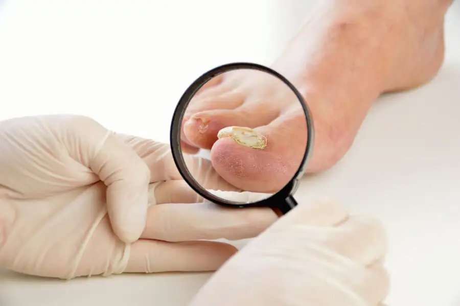
Feet (athlete’s foot)
- Itching and burning sensation,
- Cracked and dead skin (scales), often between the third and fourth toes (5)
Diagnosing SFIs using molecular analysis
Using a skin or nail sample taken by a qualified professional, molecular analysis can detect more than 50 types of fungi at the same time: dermatophytes, yeasts, and moulds, whether they are in single or mixed mycoses.
Molecular DNA detection, performed at an accredited lab, identifies each type of fungus that may be causing the SFI. This can determine the fungus/fungi causing it quickly and very precisely.
The advantages of molecular analysis
A more precise diagnosis
Diagnosing SFIs can be complex because their symptoms are often similar to the symptoms of other skin conditions such as psoriasis, eczema, autoimmune diseases, bacterial infections, and tumours.
Because it is highly sensitive and specific, molecular analysis can confirm or rule out the presence of an SFI, making it easier to apply the right treatment. As well, identifying the fungus/fungi causing the infection helps determine its source, which can prevent reinfections.
Diagnosing mixed infections
Mixed infections, especially ones caused by non-dermatophyte moulds, are often more difficult to treat. They are also associated with a higher rate of recurrence. Molecular analysis can detect multiple infectious agents from a single sample, making treatment more successful.
Reliability
Molecular analysis can detection over 50 types of dermatophytes, yeasts, and moulds at the same time, including some that are difficult, if not impossible, to identify using traditional methods such as culturing.
Fast results
Results are available 7 days after the analysis. The diagnosis is much faster and more precise than using microscopy or culturing; it can take four weeks or longer to receive those results.
Getting a specific, reliable diagnosis
Our services can help your podiatrist diagnose SFIs of the nails and skin, and determine the right treatment.
References
1. Sai BS, Tejashree A, Veeranna S, Krishna Karthik M. Speciation and in vitro activity of four antifungal drugs against clinical isolates of dermatophytes by E-test method. Int J Sci Res. 2019;8.
2. Ameli. Available [in French only] from: https://www.ameli.fr/assure/sante/themes/mycose-cutanee-peau/prevention.
3. Chris G. Adigun. Merck Manual. Available from: https://www.merckmanuals.com/en-ca/home/skin-disorders/nail-disorders/onychomycosis.
4. [in French only] https://publications.msss.gouv.qc.ca/msss/fichiers/guide-garderie/chap7-tinea-capitis.pdf
5. https://www.msdmanuals.com/home/skin-disorders/fungal-skin-infections/athlete-s-foot-tinea-pedis
6. [in French only] https://dermato-info.fr/fr/les-maladies-de-la-peau/les-mycoses-cutan%C3%A9es#mycose_description
7. Salakshna N, Bunyaratavej S, Matthapan L, Lertrujiwanit K, Leeyaphan C. A cohort study of risk factors, clinical presentations and outcomes for dermatophyte, nondermatophyte, and mixed toenail infections. J Am Acad Dermatol. 2018;79:1145–1146.
8. Antuori A, Fernández G, Fernández A, Alcaide M, Boada A, Bielsa MI, Romaní N, Matas L. Epidemiology of dermatophytic infections between 2008 and 2017 in Barcelona, Spain. Enferm Infecc Microbiol Clin. 2019;37:642–647.
9. https://cps.ca/en/documents/position/antifungal-agents-common-infections
10. Salakshna N, Bunyaratavej S, Matthapan L, Lertrujiwanit K, Leeyaphan C. A cohort study of risk factors, clinical presentations and outcomes for dermatophyte, nondermatophyte, and mixed toenail infections. J Am Acad Dermatol. 2018;79:1145–1146.